
Subungual exostosis
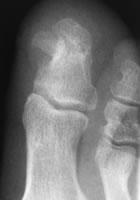
True subungual exostoses arise from the tuft of the distal phalanx. They are composed of mature bone with a fibrocartilaginous cap. Lee et al (2007) noted that half of their subungual lesions were actually osteochondromas arising from the proximal part of the distal phalanx and covered with hyaline cartilage organised as in a growth plate. True exostoses are commonest in young adults with a female predominance. They mainly occur in the great toe, although they also occur in the lesser toes and fingers.
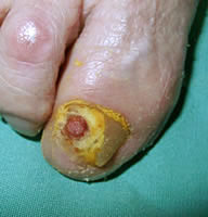
They present with a complaint of pain in the toe, sometimes localised to the nail fold. Sometimes the swelling itself may cause pressure on the shoe. Some have a history of trauma or previous nailbed surgery (which may have been for an "ingrowing toenail' that was, in fact, the exostosis).
Examination shows a firm swelling under the nail, usually in the medial nail fold. It is usually covered with epidermis, but may be raw or granulating.
The main differential diagnosis is ingrowing toenail, with a nailbed tumour such as melanoma, squamous carcinoma or glomus tumour as a much rarer possibility.
The lesion normally continues to grow so is best removed when diagnosed. This can be done under digital block anaesthesia as a day case. Sometimes the nail fold can be elevated and preserved, but usually it cannot be separated from the lesion and must be sacrificed. Even with careful excision of the whole lesion, the recurrence rate averages about 10%. A few patients require removal of so much nail bed that there is significant post-operative nail deformity, so that plastic nail bed reconstruction may be consdered (Suga 2005).
References
- De Berker DA, Langtry J. (1999). Treatment of subungual exostoses by elective day case surgery. Br J Dermatol 140(5): 915-8
- Dalle S e al. Squamous cell carcinoma of the nail apparatus: clinicopathological study of 35 cases. Br J Dermatol. 2007;156(5):871-4
- Gray RJ et al. Diagnosis and treatment of malignant melanoma of the foot. Foot Ankle Int 2006; 27:696-705
- Lee SK et al. Two distinctive subungual pathologies. Subungual exostosis and subungual osteochondroma. Foot Ankle Int 2007; 28:595-601
- Suga H et al. Subungual exostosis: a review of 16 cases focusing on postoperative deformity of the nail. Ann Plast Surg. 2005;55(3):272-5